echo lv segments | left ventricular segmentation diagram echo lv segments assembled by the American Society of Echocardiography and the European Association of Cardiovascular Imaging. This document provides updated normal values for all four cardiac . Large model, hand-wound mechanical movement, rose gold, diamonds, leather. Discover the full Tank Watch Collection on the Official Cartier® Online US Store. Elegance that has captivated the world's most astute minds.
0 · what is fractional shortening echo
1 · normal lv size and function
2 · lv wall thickness echo
3 · lv wall segments echo
4 · left ventricular segmentation diagram
5 · how to assess lv function
6 · 17 wall segments echo
7 · 17 segments of the heart
The Griffin House, built circa 1827, stands on 50 acres of land situated on .
Standardized myocardial segmentation and nomenclature for echocardiography. The left ventricle is divided into 17 segments for 2D echocardiography. One .Assessment of left ventricular systolic function has a central role in the evaluation of cardiac disease. Accurate assessment is essential to guide management .
Although certain variability exists in the coronary artery blood supply to myocardial segments, segments are usually attributed to the three major coronary arteries. Visual Assessment Semi .assembled by the American Society of Echocardiography and the European Association of Cardiovascular Imaging. This document provides updated normal values for all four cardiac . preload: end-diastolic volume (if low think -> hypovolaemia, low SVR, severe AR or MR, VSD) afterload: end-systolic wall stress (rarely used in clinical practice) LV wall thickness: .Complete review on left ventricular systolic function, with emphasis on echocardiography, definitions, methods and guidelines.
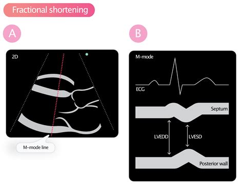
2-dimensional (2-D) echocardiography (Figure 2A) can be performed in the para-sternal long- and short-axis views by placing the calipers perpendicular to the ventricular long axis. Change in .Standardized myocardial segmentation and nomenclature for echocardiography. The left ventricle is divided into 17 segments for 2D echocardiography. One can identify these segments in multiple views. The basal part is divided into six segments of 60° each.Assessment of left ventricular systolic function has a central role in the evaluation of cardiac disease. Accurate assessment is essential to guide management and prognosis. Numerous echocardiographic techniques are used in the assessment, each .
Although certain variability exists in the coronary artery blood supply to myocardial segments, segments are usually attributed to the three major coronary arteries. Visual Assessment Semi quantitative wall motion score (1-4) can be assigned to each segment to .assembled by the American Society of Echocardiography and the European Association of Cardiovascular Imaging. This document provides updated normal values for all four cardiac chambers, including three-dimensional echocardiography and myocardial deformation, when possible, on the basis of considerably preload: end-diastolic volume (if low think -> hypovolaemia, low SVR, severe AR or MR, VSD) afterload: end-systolic wall stress (rarely used in clinical practice) LV wall thickness: > 1.5cm = LVH, < 0.6cm = LV thinning. Regional Function. 16 segments. contractility: grades.

what is fractional shortening echo
Complete review on left ventricular systolic function, with emphasis on echocardiography, definitions, methods and guidelines.2-dimensional (2-D) echocardiography (Figure 2A) can be performed in the para-sternal long- and short-axis views by placing the calipers perpendicular to the ventricular long axis. Change in LV cavity dimensions during systole can be used to calculate .
A normal LV ejection fraction in the presence of the heart failure syndrome leads to a search for diastolic dysfunction. Typical echo findings in diastolic dysfunction are normal LV cavity size, thickened ventricle, and reversed E/A ratio.
Echocardiography is the principal modality for investigating left ventricular systolic function and diastolic function. M-mode, 2D echocardiography and Doppler are all used to examine various parameters.
This chapter demonstrates left chamber quantification through various measurements of left ventricular size and dimensions, left ventricular mass, left ventricularglobal function, regional wall motion, left ventricular segmentation, global left ventricular .
Standardized myocardial segmentation and nomenclature for echocardiography. The left ventricle is divided into 17 segments for 2D echocardiography. One can identify these segments in multiple views. The basal part is divided into six segments of 60° each.Assessment of left ventricular systolic function has a central role in the evaluation of cardiac disease. Accurate assessment is essential to guide management and prognosis. Numerous echocardiographic techniques are used in the assessment, each .Although certain variability exists in the coronary artery blood supply to myocardial segments, segments are usually attributed to the three major coronary arteries. Visual Assessment Semi quantitative wall motion score (1-4) can be assigned to each segment to .
assembled by the American Society of Echocardiography and the European Association of Cardiovascular Imaging. This document provides updated normal values for all four cardiac chambers, including three-dimensional echocardiography and myocardial deformation, when possible, on the basis of considerably preload: end-diastolic volume (if low think -> hypovolaemia, low SVR, severe AR or MR, VSD) afterload: end-systolic wall stress (rarely used in clinical practice) LV wall thickness: > 1.5cm = LVH, < 0.6cm = LV thinning. Regional Function. 16 segments. contractility: grades.Complete review on left ventricular systolic function, with emphasis on echocardiography, definitions, methods and guidelines.
2-dimensional (2-D) echocardiography (Figure 2A) can be performed in the para-sternal long- and short-axis views by placing the calipers perpendicular to the ventricular long axis. Change in LV cavity dimensions during systole can be used to calculate .A normal LV ejection fraction in the presence of the heart failure syndrome leads to a search for diastolic dysfunction. Typical echo findings in diastolic dysfunction are normal LV cavity size, thickened ventricle, and reversed E/A ratio.Echocardiography is the principal modality for investigating left ventricular systolic function and diastolic function. M-mode, 2D echocardiography and Doppler are all used to examine various parameters.
normal lv size and function
ciabatte gucci uomp
cinto gucci amazon
cinta elastico gucci
lv wall thickness echo
The Italian Man Who Went To Malta is a parody video created by Jokhie Judy and uploaded to YouTube on October 8, 2005. This video features the Italian accent made by Alundras on YouTube. The video revolves .
echo lv segments|left ventricular segmentation diagram


